This week I’d like to start a series of blogs relating to valvular heart disease, starting with a description of the heart valves and how they work. We have four valves in our hearts and their purpose is to keep blood moving in a single direction. They open during one phase of the cardiac cycle (systole or diastole, depending on which valve it is), allowing blood to pass from one chamber to another; and they close during the other phase, preventing blood from leaking back or regurgitating.
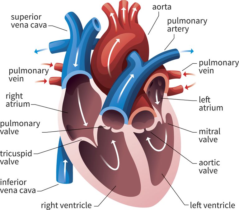
Two of the valves—the aortic and the mitral—sit in the left side of the heart; two—the pulmonic and the tricuspid—sit in the right side of the heart. The aortic and pulmonic (or pulmonary) valves are the gateways between the ventricles and the great vessels. The aortic sits between the left ventricle and the aorta, the large vessel that supplies the body with blood. The pulmonic sits between the right ventricle and the pulmonary artery, the large vessel that supplies the lungs with blood. The mitral valve sits between the left atrium and left ventricle and the tricuspid valve between the right atrium and right ventricle. Below is an image of these valves and the pathway of blood flow or circulation.

Implicit in this discussion is that we have two parallel circulations—one for the lungs and one for the body. We call these the pulmonary and systemic circulations, respectively. These circulations are connected, in that the blood from the lungs is collected in the pulmonary veins and returned to the left side of the heart (the left atrium), while the blood from the body flows through the superior and inferior vena cavae (plural of cava) into the right side of the heart (the right atrium).
Two main problems can occur with valves—they can develop stenosis, which means that they don’t open all the way. Or they can develop regurgitation, which means they don’t close all the way. You might recall the word “stenosis” in an earlier discussion of “coronary stenosis.” Stenosis is a term we use with various heart or vascular problems that have in common that a passageway is narrowed. While coronary stenoses are narrowings that restrict blood flow through the coronary arteries, valvular stenosis means the valve is restricted in its opening, making it more difficult for blood to get across the valve from one cardiac chamber to another.
Regurgitation of a valve is often referred to by physicians as a “leaky valve.” As with other non-medical terms that physicians use to simplify things, it can lead to some confusion. People imagine that blood must be leaking outside the heart—but it just means that when the valve closes, there is a residual opening, allowing a trickle (or a torrent, if the regurgitation is severe) of blood to flow back into the chamber from which it just came. This makes the heart have to work harder to get blood to the body (or lungs), as it must pump an extra amount to compensate for what has leaked backward.
Now that you understand the circulation of blood and how the heart valves are supposed to work, we’ll turn in the next several blogs to the types of valvular heart disease that are most common.
Greg Koshkarian, MD, FACC
